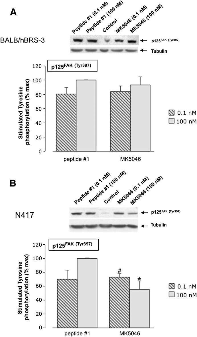
Fig. 9.
Peptide #1 and MK-5046 stimulate Tyr397 phosphorylation of FAK in (A) hBRS-3-transfected Balb 3T3 cells and (B) NCI-N417 native hBRS-3 cells. Above each panel are representative Western blots of the stimulation of p125FAK(Tyr397) in Balb 3T3 cells (A) and NCI-N417 cells (B) treated with 0.1 nM and 100 nM of peptide #1 or MK-5046 and incubated for 3 minutes at 37°C. Each panel shows the mean ± S.E.M. from at least four experiments. The results are expressed as the percentage of the maximal concentration of peptide #1 (100 nM). *P < 0.05 versus 100 nM peptide #1. #P < 0.05 versus 100 nM MK-5046.

