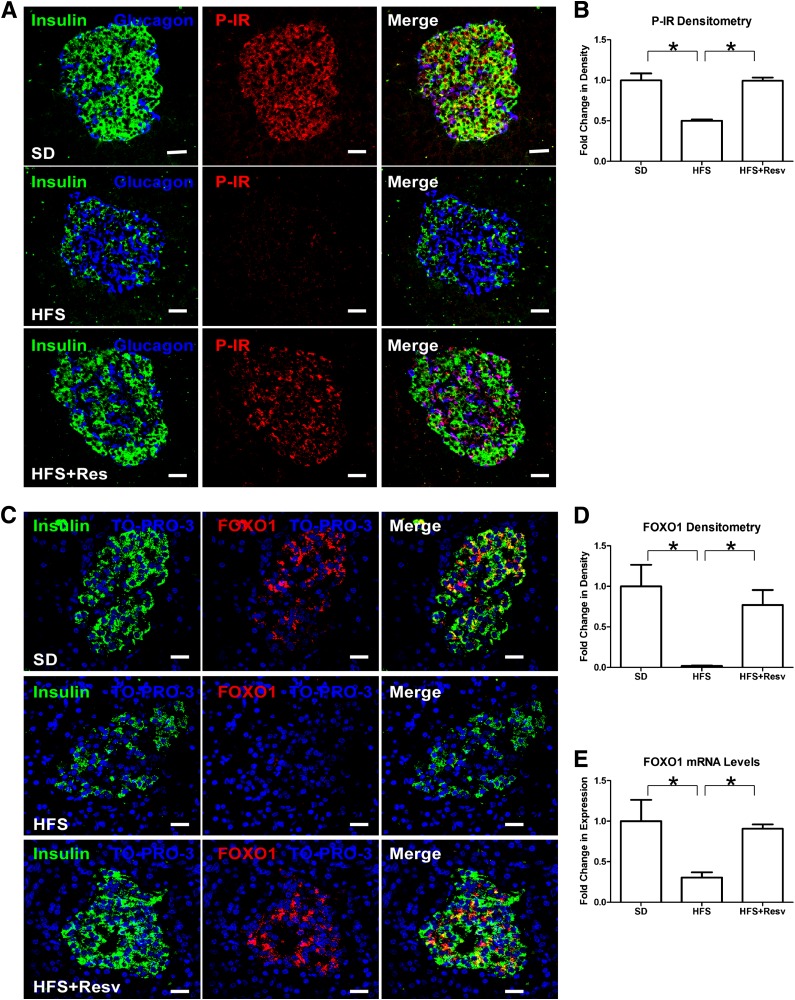
FIG. 3.
Decreased P-IR and total FOXO1 levels in an HFS diet. A: Immunostaining for insulin (green), glucagon (blue), and tyrosine P-IR (red) in islets of monkeys after 24 months on SD (top panel), HFS diet (middle panel), or HFS+Resv diet (bottom panel). Scale bar = 20 µm. B: Quantitation of signal intensity for P-IR. Data are shown as the mean ± SEM. *P < 0.05. C: Immunostaining for insulin (green), TO-PRO-3 (blue), and total FOXO1 (red) in islets of monkeys after 24 months on SD (top panel), HFS (middle panel), or HFS+Resv (bottom panel) diets. Scale bar = 20 µm. D and E: Quantitation of signal intensity (D) and mRNA levels (E) for FOXO1. Data are shown as the mean ± SEM. *P < 0.05.

