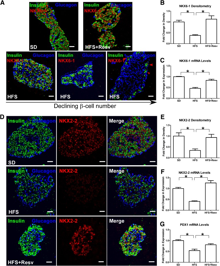
FIG. 4.
Decreased nuclear expression of NKX6–1, NKX2–2, and PDX1 after an HFS diet. A: Immunostaining for insulin (green), glucagon (blue), and NKX6–1 (red) in monkeys after 24 months on the indicated diet. The islets from different HFS monkeys are shown in order of declining β-cell numbers (bottom panel); the red arrows indicate depletion of nuclear NKX6–1 in insulin-expressing cells. Scale bar = 20 µm. B and C: Quantitation of signal intensity (B) and mRNA levels (C) for NKX6–1. Data are shown as the mean ± SEM. *P < 0.05. D: Immunostaining for insulin (green), glucagon (blue), and NKX2–2 (red) in islets of monkeys after 24 months on SD (top panel), HFS diet (middle panel), or HFS+Resv diet (bottom panel). Scale bar = 20 µm. E and F: Quantitation of signal intensity (E) and mRNA levels (F) for NKX2–2. Data are shown as the mean ± SEM. *P < 0.05. G: Quantitation of mRNA levels for PDX1. Data are shown as the mean ± SEM. *P < 0.05.

