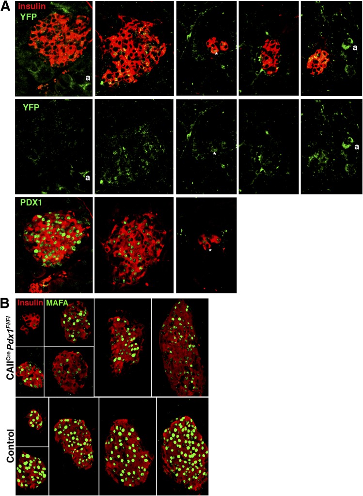
FIG. 6.
Islets with PDX1null β-cells show lineage tracing marker and low to undetectable MAFA expression. A: The variation of PDX1 immunostaining corresponded with the expression of lineage marker YFP in islets from a 4-week-old CAIICre;Pdx1Fl/Fl (blood glucose: 278 mg/dL) mouse. The middle panel shows YFP expression as split green channel of images shown in the top panel (insulin, red; YFP, green). The bottom panel shows same islets on adjacent section (due to antibody compatibility issues) with PDX1 (green) and insulin (red). a, lineage-marked acinar cell. *Identifies the same cell in different images. B: MAFA expression (green) showed similar variation from high intensity to low/undetectable in insulin+ (red) islets from same section of a 10-week-old CAIICre;Pdx1Fl/Fl mouse (blood glucose at 4 weeks: 272 mg/dL, 10 weeks: 189 mg/dL) compared with homogeneous high intensity of control littermate (blood glucose at 4 weeks: 172 mg/dL, 10 weeks: 178 mg/dL).

