FIG. 5.
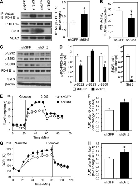
Sirt3 knockdown in C2C12 myoblasts impairs PDH activity despite decreased phosphorylation of PDH E1α and leads to a substrate switch toward fatty acid utilization. A: Total mitochondrial protein lysates from shGFP control and shSirt3 myoblasts were immunoprecipitated (IP) with AcK (AcLys) antibody and subjected to Western blot analysis (IB) using an anti–PDH E1α antibody. The same mitochondrial lysates were directly subjected to Western blot analysis using antibodies against PDH E1α, Sirt3, and voltage-dependent anion channel (VDAC) as a mitochondrial loading control. Densitometry of PDH E1α from AcK immunoprecipitates was normalized to total PDH E1α (n = 4 separate experiments; *P < 0.05, Student t test). B: Total PDH activity was assessed in confluent control and shSirt3 myoblasts using PDH activity microplate assay kit and normalized to total protein from detergent extraction (n = 5 separate experiments; †P < 0.05, paired t test). C: Phosphorylation of PDH E1α and total Sirt3 levels were determined by Western blot analysis of whole cell lysates from confluent shGFP and shSirt3 C2C12 myoblasts. D: Densitometry of Western blots from C (n = 3 separate experiments, *P < 0.05, Student t test). E: ECAR was measured in shSirt3 and control myoblasts using a Seahorse flux analyzer after incubation in glucose-free Seahorse running media for 1 h at 37°C. A representative tracing of basal and glucose-stimulated ECARs recorded before and after addition of 25 mmol/L glucose (final concentration) is shown. At the end of the glucose metabolism period, 2-deoxyglucose was injected to give a final concentration of 25 mmol/L. F: AUC calculation of glucose-stimulated ECAR from E. G: A representative tracing of palmitate OCR measured in control and Sirt3 knockdown cells after incubation with substrate-free buffer for 1 h at 37°C. Basal and palmitate-BSA–stimulated OCRs were recorded and plotted as a percentage over basal OCR. Finally, 50 μmol/L etomoxir was injected. H: AUC of palmitate-stimulated OCR from G (n = 3; *P < 0.05, Student t test). mOD, milli optical density.

