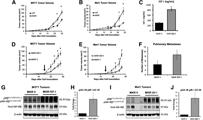
FIG. 2.
IGF-I increased orthotopic MVT1 and Met1 tumor growth in the hyperinsulinemic MKR mice by increasing IGF-IR phosphorylation. WT and MKR mice were injected with tumor cells on Day 0. MKR mice developed larger MVT1 and Met1 tumors than WT mice (A and B). MVT1 and Met1 tumor cells were orthotopically injected into MKR mice, mice were divided into two groups with equal mean tumor size, and mice were administered either rhIGF-I or vehicle (vertical arrow indicates time when treatment began). Administration of rhIGF-I led to a further stimulation in tumor growth, over endogenous hyperinsulinemia (D and E). Serum IGF-I concentration in the rhIGF-I treatment group was 2.6 times that of the control group (C). The greater mean number of pulmonary macrometastases in the mice treated with rhIGF-I did not reach statistical significance, compared with vehicle-treated MKR mice (F). Western blot analysis of tumor lysates demonstrated that MKR mice with endogenous hyperinsulinemia (MKR V) show IRβ phosphorylation at 95 kDa, and rhIGF-I treatment led to increased IGF-IRβ and IRβ or hybrid receptor phosphorylation in MVT1 and Met1 tumors (G–J). Representative images from three repeated experiments of tumor volume and Western blots are displayed. The graphs represent the average for each group; error bars indicate SEM (A–F, H, and I). Statistical analysis was performed using a two-tailed t test; *indicates statistically significant differences (P < 0.05) between the groups. n = 8–11 mice per group. p, phosphorylated.

