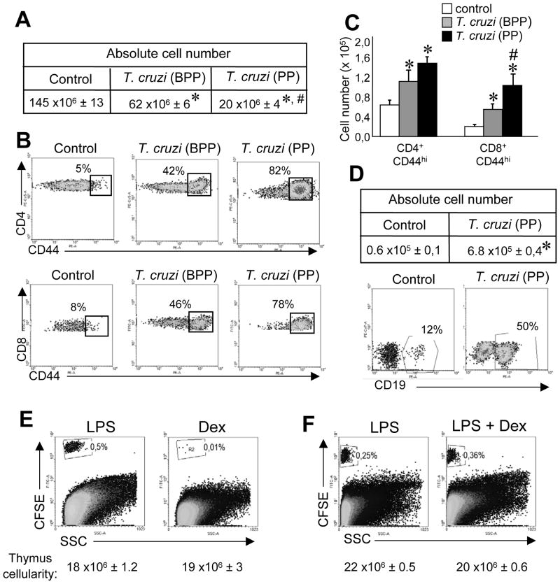
Figure 2. Thymic cell loss is necessary but it is not the only requirement to permit the ingress of peripheral cells into the thymus.
Mice infected with 5 × 10⁵ trypomastigotes (i.p) were killed the day of parasitemia peak (PP) or before the day of parasitemia peak (BPP). (A) Total cell number or (B) the percentages and (C) absolute cell numbers of CD4+ CD44+ or CD8+ CD44+ cells at both times. (D) The absolute cell number and the percentage of B cells (CD19+) in the thymic DN compartment were determined in control mice and in T. cruzi-infected mice sacrificed during PP. (E) Percentage of CFSE+ cells in the thymi of LPS treated mice (see the Materials and methods) versus mice treated with a single dose of 0.3 mg of dexamethasone (Dex). (F) Percentage of CFSE+ cells in the thymi of LPS treated mice versus mice treated with LPS and a single dose of 0.3 mg of Dex. Data from B and D was obtained by gating on the viable cells from thymi of control or T. cruzi-infected mice and later on the CD4+, CD8+ or DN cells as shown in Supporting Information fig. 3B. Data from A, C and D are shown as the mean + SEM of 18 mice per group pooled from 6 independent experiments. (B, D, E and F) Representative dot plots from individual recipient mice are shown. Data are representative of at least 3 independent experiments with 2–3 mice per group. *Control vs T. cruzi-infected, p<0.05, #T. cruzi-BPP vs T. cruzi-PP, p<0.05. Student unpaired t test.

