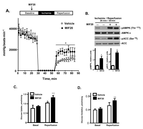
Figure 4.
MIF20 stimulates cardiac AMPK activation and glucose uptake, and ameliorates post-ischemic cardiac dysfunction. Isolated hearts from C57BL/6 WT mice underwent 30 minutes of baseline perfusion, 25 minutes of global no-flow ischemia, and 30 minutes of reperfusion. The MIF agonist, MIF20, was added to the perfusion buffer during baseline perfusion 15 min prior to ischemia at a final concentration of 8 nM. (A) Heart rate-left-ventricular developed pressure product during baseline and post-ischemic reperfusion (n=4 hearts for each group. Mean ± SEM, *p<0.05 vs. vehicle controls). (B) Representative western blots for phosphorylated AMPK and ACC in cardiac tissue from two hearts during reperfusion, and after treatment with vehicle or MIF20. Tissue lysates were prepared by freeze-clamping cardiac muscle 30 mins after initiation of reperfusion. The densitometric analysis is for 4 hearts per group (Mean ± SEM, *p<0.05 vs. vehicle). (C) Measurement of glucose uptake in cardiac tissue during baseline and post-ischemic reperfusion periods after treatment with vehicle or MIF20. The perfusate was collected at 1, 5, and 10 mins after beginning reperfusion and the values averaged for each heart. (n=4 hearts for each group. Mean + SEM, *p<0.05 for reperfusion vs. corresponding baseline; †p<0.05 for MIF20 vs. vehicle). (D) Enhancement of MIF-dependent glucose oxidation rate by MIF20 (8 nM) in a separate group of anterograde perfused working mouse hearts subjected to 15 min ischemia and 30 mins reperfusion (n=4 hearts per experimental group; *p<0.05 vs. corresponding baseline; †p<0.05 for MIF20 vs. vehicle).

