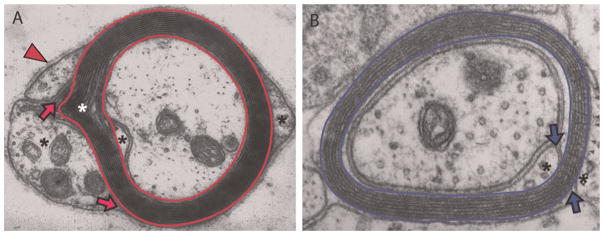
Fig. 1. Electron micrographs of rodent (A) PNS and (B) CNS myelin sheaths (originals kindly provided by Dr. Jack Rosenbluth, New York University School of Medicine).

Compact myelin (bordered by red bands in PNS and by blue bands in CNS) is surrounded by glial cell cytoplasm (*), containing organelles where myelin-destined lipids and proteins are synthesized. For peripheral nerves, a supportive basement membrane (arrowhead) is also present and functions to stabilize the fibers and signal myelination (Jessen and Mirsky, 2005). Regions of compact myelin, where MBP, MPZ and PLP1 reside, are separated from regions of non-compacted myelin where MBP, MPZ and PLP1 are made, by tight junctions that form inner and outer mesaxons (arrows). Of many proteins synthesized in glial cell cytoplasm, few accumulate in and help structure myelin. A Schmidt–Lanterman incisure, with cytoplasm included between layers is common in MPZ-based though not PLP1-based myelin (
 ).
).
