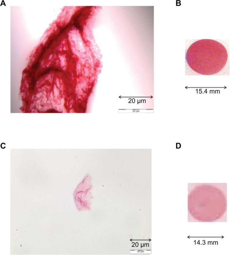
Figure 3.

Detection of 1 μL of HSA (200 mg/mL) dotted onto NC membrane after incubation in GCC (10 mL), including different amounts (B, 10 μL; D, 1 μL) of labeled mc-anti-HSA Ab. Images of these GCC-labeled Ab solutions (A, 10 μL; C, 1 μL) were checked by microscopy (Olympus).
