
Figure 2.
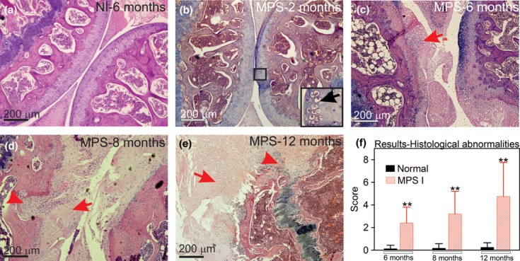
Aspect of knee joints using H–E stain. (a) Joint sections representative from a wt mouse at 6 months; (b) MPS I mice at 2 months, insert indicates GAG storage; (c) MPS I mouse at 6 months, with the arrow indicating damage to the articular surface; (d) MPS I mouse at 8 months, with the arrowhead representing bone resorption and the arrow indicating fibrocartilaginous proliferation; (e) MPS I mouse at 12 months, with the arrowhead showing intense bone resorption and the arrow indicating intense fibrocartilaginous proliferation; and (f) results from histological scores obtained in wt and MPS I mice joints at 6, 8 and 12 months (N = 6–11). Score was based on abnormalities described in Figure 1 and detailed in the methods section, where the higher the score, the worse the joint. The maximum score is 10. **P < 0.01, using Student's t-test. N = 6–11 mice/group.