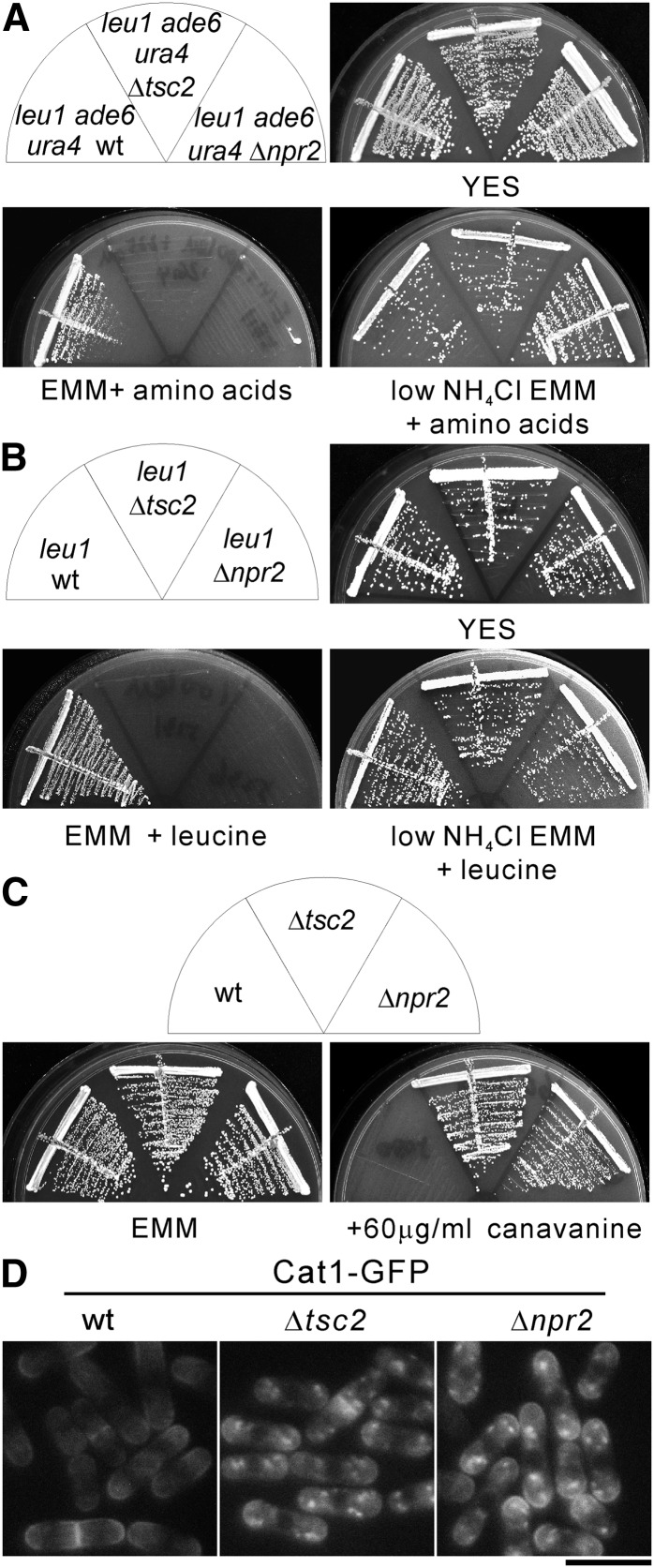
Figure 1.
The Δnpr2 and Δtsc2 cells display similar phenotypes. (A) The wild-type (KP93006), Δtsc2 (KP91087), and Δnpr2 (KP90390) cells auxotrophic for leucine, adenine, and uracil were streaked onto YES, EMM (5 g/liter NH4Cl), or low NH4Cl EMM (0.5 g/liter NH4Cl) supplemented with 100 mg/liter leucine, 225 mg/liter adenine, and 225 mg/liter uracil. The plates were incubated at 27° for 3 days (YES), 4 days (low NH4Cl EMM + amino acids), or 5 days (EMM + amino acids), respectively. (B) The leucine auxotrophic cells of wild-type (HM123), Δtsc2 (KP5131), and Δnpr2 (KP5236) were streaked onto YES, EMM (5 g/liter NH4Cl), or low NH4Cl EMM (0.5 g/liter NH4Cl) supplemented with 100 mg/liter leucine. The plates were incubated as described in Figure 1A. (C) The prototrophic cells of wild-type (KP5080), Δtsc2 (KP5128), and Δnpr2 (KP5237) were streaked onto EMM or EMM containing 60 μg/ml canavanine. The plates were incubated at 27° for 4 days (EMM) or 5 days (+60 μg/ml canavanine). (D) Subcellular localization of Cat1 in wild-type, Δtsc2, and Δnpr2 cells. The wild-type (KP5859), Δtsc2 (KP5826), and Δnpr2 (KP5822) cells expressing chromosome-borne Cat1–GFP under the control of its own promoter were grown to early log phase in EMM media at 27°. The fluorescence of the Cat1–GFP was examined. Bar, 10 μm.

