
Figure 3.
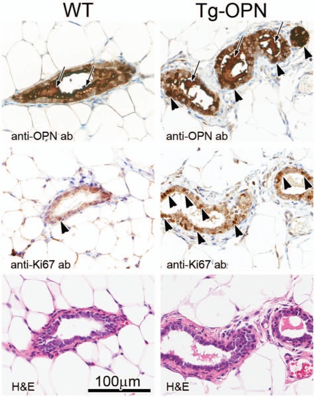
Mammary gland histology, osteopontin (OPN) expression, and proliferation in nulliparous 10-week female transgenic (Tg)-OPN versus wild type (WT) control mice. WT (left) and Tg-OPN (right) mammary ducts, serial or near serial sections. Immunohistochemistry against murine OPN (top panels) shows the abundant expression of OPN in normal mammary glands with secretion into the ductal lumen (arrows). In the OPN transgenic gland, there is luminal secretion (arrows) as well as cytoplasmic excess accumulation (arrowheads). Proliferation as assessed by immunohistochemistry for Ki67 (middle panels) reveals areas with proliferation exceeding any seen in wild type mice. In the representative areas there is one positive nucleus in the wild type (arrowhead), whereas in the Tg-OPN there are many positive nuclei (arrowheads mark only some of the positive cell nuclei). Hematoxylin-eosin (H&E) stained serial sections are shown below, with the 100 µm scale bar (bottom left) pertaining to all panels.