
Figure 4.
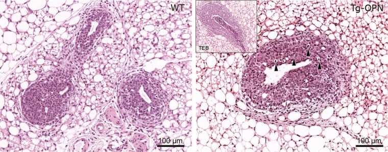
Osteopontin (OPN)-induced epithelial hyperplasia in young nulliparous mammary gland. Tissue sections (4 µm) of formalin-fixed, paraffin-embedded abdominal (#4) mammary glands from 4-week-old age-matched nulliparous female wild type (panel WT) and transgenic osteopontin (Tg-OPN) littermates were stained with hematoxylin-eosin. Tg-OPN induced lobular hyperplasia with increased apoptosis (arrowheads, Tg-OPN panel). Inset in Tg-OPN panel shows a representative Tg-OPN terminal end bud (TEB) with cell debris in lumen (L).