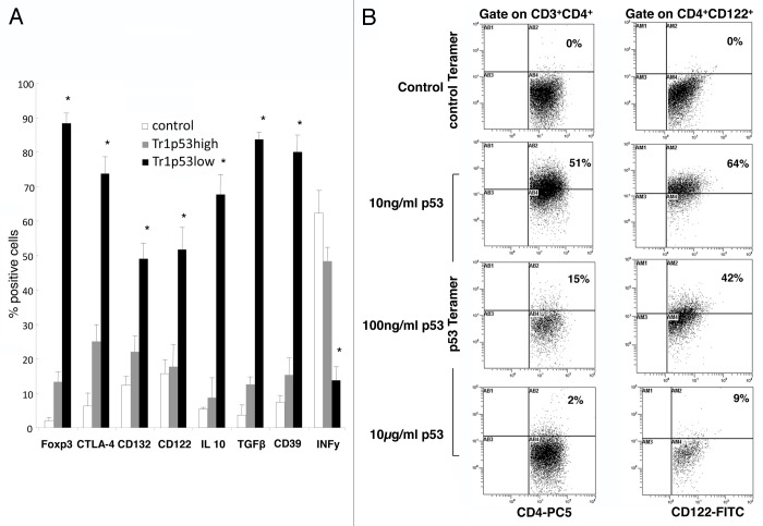
Figure 2. Phenotype and antigen specificity of p53-specific Tr1 cells generated in vitro. (A) Cytofluorometric analysis of Tr1 cells generated in the presence of autologous immature dendritic cells (iDCs) pulsed with various doses of a wild-type p53-derived (p53108–122) peptide. CD4+CD25− T cells cultured for 10 d in the presence of 150 IU/mL interleukin-2 and unpulsed iDCs served as control cells. Data are means ± SD (n = 5 independent experiments); *P < 0.05. (B) Tr1 cells were stained with a control or a p53108–122-specific tetramer, followed by the quantification of tetramer+ T cells by flow cytometry. One representative experiment out of 5 performed with cells from different donors is shown.

An official website of the United States government
Here's how you know
Official websites use .gov
A
.gov website belongs to an official
government organization in the United States.
Secure .gov websites use HTTPS
A lock (
) or https:// means you've safely
connected to the .gov website. Share sensitive
information only on official, secure websites.
