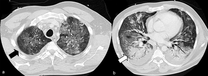
Figure 2.

IV contrast-enhanced chest CT scan (lung window) in a 47-year-old male patient with influenza provoked acute respiratory distress syndrome (ARDS). Ground glass–type increases in density (a, black arrow) and areas of consolidation (b, white arrow) are recognizable. Reprinted from: Grieser C, Goldmann A, Steffen I et al.: Computed tomography findings from patients with ARDS due to Influenza A (H1N1) virus-associated pneumonia. Eur J Radiol 2012; 81: 389–94. Used by permission of the publisher (Elsevier)
