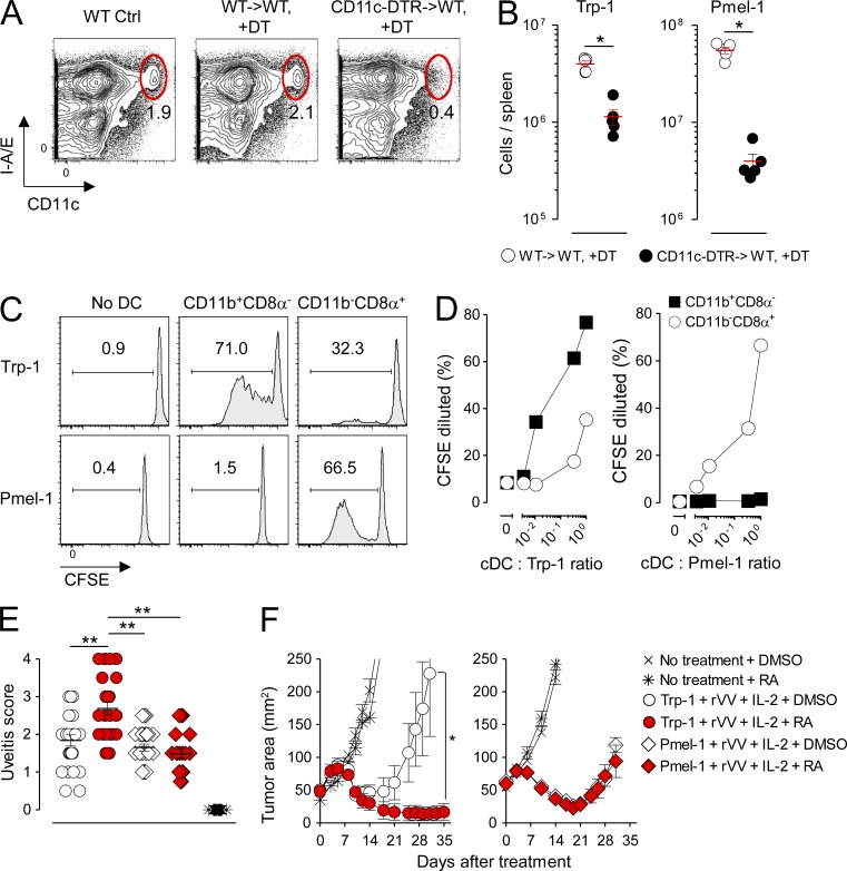
Figure 10.
RA selectively augments MHC class II– but not class I–restricted autoimmunity and antitumor immunity in mice rendered acutely VAD through TBI conditioning. Lethally irradiated (9 Gy) mice were reconstituted either with WT BM (WT→WT) or BM derived from CD11c–diphtheria toxin receptor mice (CD11c-DTR→WT). After reconstitution, radiation chimeras were treated with diphtheria toxin (DT), and 106 naive Trp-1 CD4+ T cells or Pmel-1 CD8+ T cells were adoptively transferred in combination with 2 × 10⁷ PFU of an rVV encoding cognate Ag for the transferred T cells. (A and B) Representative FACS plots showing depletion of splenic cDCs (CD11chighI-A/E+; A) and scatter plots showing the absolute number of transferred Trp-1 CD4+ T cells or Pmel-1 CD8+ T cells in the spleen at the peak (day 5) of the immune response after vaccination (B); n = 4 mice per group. This experiment was repeated twice with similar results. (C and D) Representative FACS plots showing the proliferation of naive, Ly5.2+ CFSE-labeled Trp-1 CD4+ or Pmel-1 CD8+ T cells in response to stimulation with FACS-sorted Ly5.1+ splenic CD11b+CD8α− and CD11b−CD8α+ cDC subsets isolated 36 h after in vivo vaccination with rVV at a 1:1 ratio (C) or at titrated cDC/T cell ratios (D). CFSE proliferation was measured on day 5 after stimulation after gating on live+, singlet+, Ly5.1−, Ly.5.2+, CD3+CD4+, or CD3+CD8+ cells. Data shown are representative of two independently performed experiments. (E) Ocular autoimmune pathology scores 14 d after mice were rendered acutely VAD in response to TBI conditioning followed by treatment with exogenous RA or DMSO Ctrl administered alone or in combination with transfer of 106 Trp-1 CD4+ or Pmel-1 CD8+ T cells. Pooled results from two independently performed experiments are shown; n = 20–30 evaluated eyes per treatment group. Horizontal bars indicate means ± SEM of individually evaluated eyes. *, P < 0.05; **, P < 0.01 (unpaired Student’s t test). (F) Treatment of 10-d established s.c. B16 melanoma tumors in mice rendered acutely VAD in response to TBI conditioning treated with exogenous RA or DMSO Ctrl administered alone or in combination with transfer of 106 Trp-1 CD4+ or Pmel-1 CD8+ T cells; n = 5 mice per treatment group. Data shown are representative of three independently performed tumor treatment experiments. Horizontal bars indicate means ± SEM of individually evaluated mice. *, P < 0.05 (Wilcoxon rank sum). All mice treated in E and F received concurrent vaccination with rVV and exogenous IL-2.

