Figure 3.
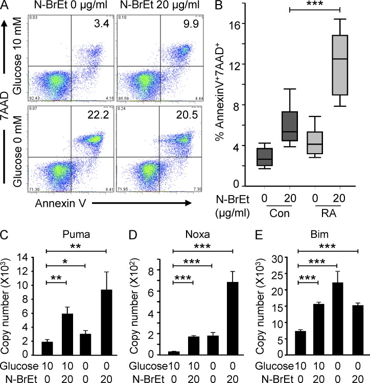
PFKFB3 deficiency renders CD4 T cells apoptosis susceptible. Naive CD4 T cells were purified and activated as in Fig. 1. On day 2, T cells were washed and treated with 20 µg/ml of the PFK-2 inhibitor N-BrEt for an additional 48 h in the absence or presence of 10 nM glucose. Apoptotic cells were detected by flow cytometry; representative data are shown (A). Inhibitor-induced increases in death rates for control and RA T cells are presented for 18 patients and 15 controls (B). Data are shown as box plots. Median, 25th, and 75th percentiles (box), and 10th and 90th percentiles (whiskers) are displayed. Transcripts of Puma (C), Noxa (D), and Bim (E) were quantified by qPCR and are shown as mean ± SEM from three independent experiments. *, P < 0.05; **, P < 0.01; ***, P < 0.001.

