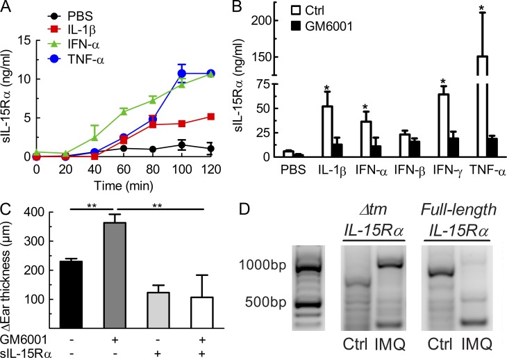
Figure 4.
Release of soluble IL-15Rα depends on proteolytic cleavage by MMPs, rather than alternative splicing of IL-15Rα. (A) Primary keratinocytes from healthy subjects were stimulated using PBS, IL-1β, IFN-α, and TNF, and supernatant was collected at different time points for measuring levels of soluble IL-15Rα (sIL-15Rα). (B) Primary human keratinocytes were treated as in A using PBS, IL-1β, IFN-α, IFN-β, IFN-γ, and TNF, without or with the MMP inhibitor GM6001, followed by determining levels of sIL-15Rα in the supernatant 2 h later. (C) WT mice received IMQ cream on their right ear for 6 consecutive days, along with intraperitoneal injections of either PBS, GM6001, sIL-15Rα, or GM6001 plus sIL-15Rα. Shown is the difference in ear thickness as in Fig. 1 C determined on day 6. (D) WT mice were treated using Vaseline (Ctrl) or IMQ on their right ear for 6 consecutive days, followed by sacrificing mice, harvesting the right ears for mRNA extraction, and performing RT-PCR for full-length IL-15Rα (around 800 bp) versus IL-15Rα lacking the transmembrane domain (Δtm) due to alternative splicing (around 500 bp). Data are representative of two to three independent experiments. P-values were determined using one-way ANOVA; *, P < 0.01; **, P < 0.001.

