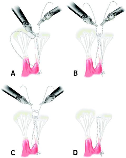
Figure 2.

Posterior neochord implantation: One arm of suture is passed through the fibrous papillary muscle tip two to three times and twice through the free edge of the prolapsed segment. The second arm is passed twice through the free edge of the prolapsed leaflet. The neochord’s length is adjusted to the height of the nearest non-prolapsed posterior leaflet segment, and the suture is tied. A, B: Figure-of-eight neochordal implantation. C: Chordal height adjustment. D: Completed neochord.
