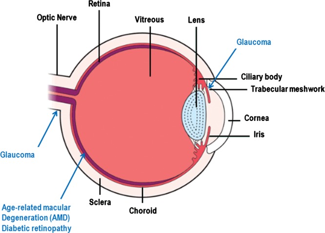
Figure 1.

Schematic diagram of the eye. The basic structures of the eye are labeled in black font and the ocular tissues affected by the disorders discussed in this review are labeled in blue font. The function of the eye is to transduce a light signal to an electrical impulse. Light enters the eye through the cornea is focused by the lens and then stimulates the photoreceptors of the retina. The photoreceptors transduce the light signal into an electrical impulse that travels through the optic nerve to the brain. AMD affects the retina, and in particular the macular region of the retina responsible for high acuity vision. The retinal pigment epithelium (also a site of damage in AMD) is the outermost layer of the retina just beneath the photoreceptors. Diabetic retinopathy also affects the retina, frequently causing damage to the macular region and also other parts of the retina. Elevated IOP in glaucoma is caused by a reduction in the rate of removal of intraocular fluid by the trabecular meshwork. The visual compromise in glaucoma is caused by damage to the optic nerve and elevated IOP is a significant risk factor for optic nerve degeneration. Myopia, or near-sightedness, is created when the image focal point occurs in front of the retina (not shown on this figure). This can occur when the eye is too long for it's inherent focusing power.
