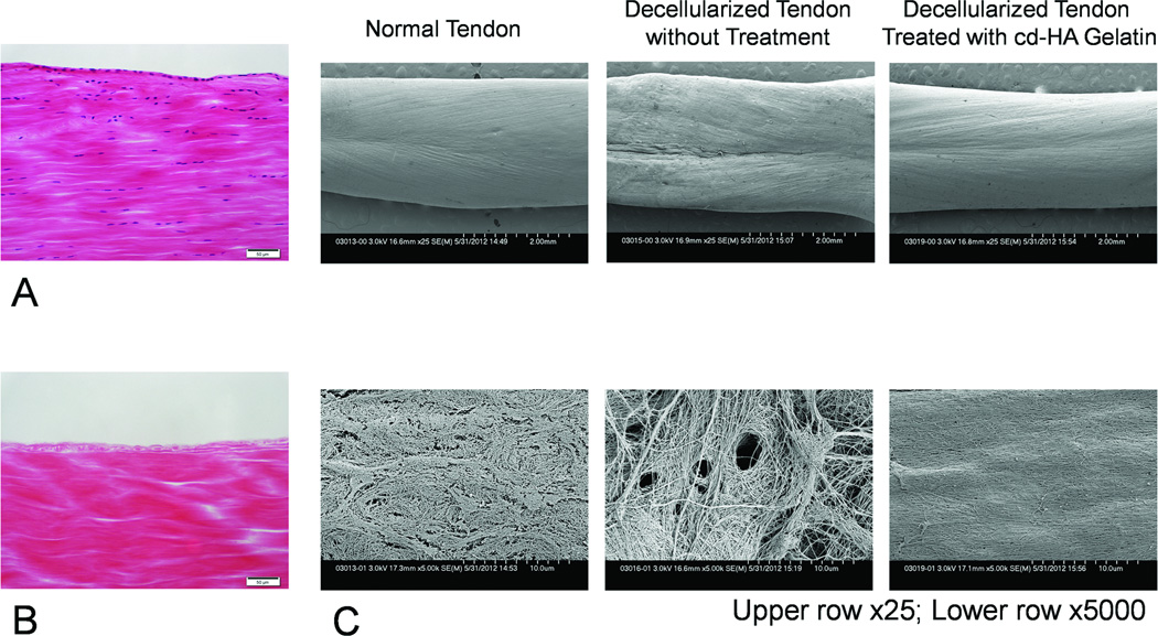
Figure 4.
Histology of longitudinal sections of normal tendon (A) and decellularized tendon (B). No cells are visible in the decellularized tendon (hematoxylin and eosin staining 200×, scale bar: 50µm). (C) Scanning electron microscopic images of the tendon after 1000 cycles of repetitive motion. Untreated, decellularized tendon surfaces appeared rough, while decellularized tendon surfaces treated with cd-HA-gelatin appeared to be smoother, and similar to normal tendon.

