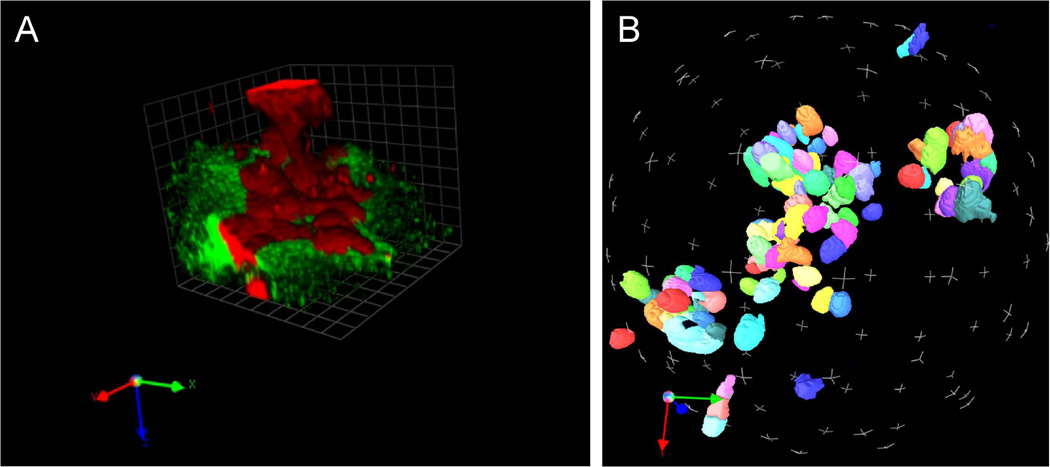
Figure 4.20.3.
Frame captures of 3-D reconstructions of human breast cancer cells grown in rBM containing DQ-collagen IV. Image stacks containing the DQ-substrate channel (green), the nuclei channel (blue) and the CellTracker channel (red) were used to create graphical 3-D reconstructions/projections using the QVTR feature of the Volocity imaging software suite. The 3-D image can be rotated 360° in order to (A) view proteolysis of DQ-substrates (green) in relation to the cells (red) or (B) view only the nuclei for the purpose of counting cells.

