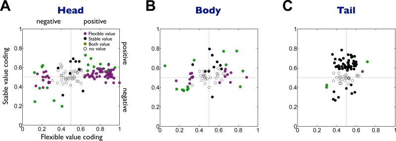
Figure 4. Flexible and stable values by individual neurons in the caudate subregions.
(A-C) Comparison between flexible value coding (abscissa) and stable value coding (ordinate) in the caudate head (n=125), body (n=65) and tail (n=87). Plotted for each neuron (each dot) are the magnitudes of the flexible and stable value coding measured by ROC areas. ROC areas higher and lower than 0.5 indicate positive and negative value coding, respectively. Purple: neurons encoding only flexible values. Black: neurons encoding only stable values. Green: neurons encoding both flexible and stable values. White: neurons encoding neither value.

