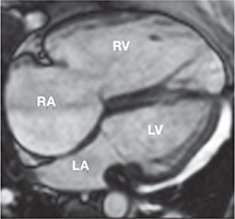
Figure 2.
Steady-state free precession four-chamber view of a patient with tetralogy of Fallot and severe pulmonary and tricuspid regurgitation with marked right atrial and right ventricular dilation. RA: right atrium; RV: right ventricle; LA: left atrium and LV: left ventricle.

