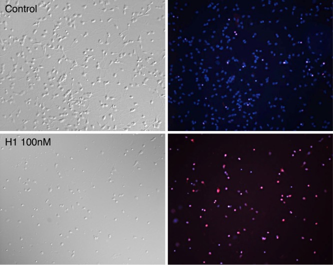
Figure 4. Neurotoxic effects of histone H1 on dissociated cortical neurons.
Cortical neurons were plated on laminin-coated coverslips for 48 hrs. Cultures were treated with histone H1 at 50, 100 or 200 nM or left as controls for a further 24 hrs when they were incubated with propidium iodide (PI) for 10 mins prior to fixing and incubation with DAPI. As propidium iodide enters dying cells and binds to nuclear DNA its red fluorescence is greatly enhanced. Top left (control) and lower left (100 nM histone H1) show phase bright examples with their corresponding DAPI (blue) and PI (red) merged images which indicate the numbers of dying cells (magenta) and total nuclei (magenta and blue).

