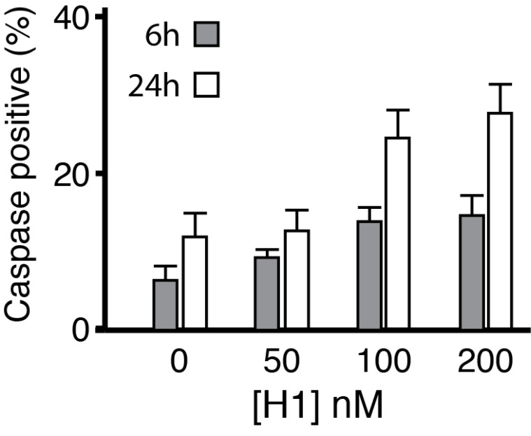
Figure 5. Histone H1 induces apoptosis.
Cortical neurons were plated on laminin-coated coverslips and treated with histone H1 at 50, 100 and 200 nM for 6 hrs and 24 hrs. Cells were fixed and stained with DAPI (total nuclei) and for activated caspase 3 to detect apoptosis. Counts for both DAPI and caspase 3 were made for between 11–14 random fields per coverslip for each condition using Image J (University of California San Francisco UCSF) and expressed as % caspase 3/DAPI. Histone H1 induced significant upregulation of activated caspase 3 at 6 and 24 hrs for neurons treated at 100 and 200 nM respectively (ANOVA).

