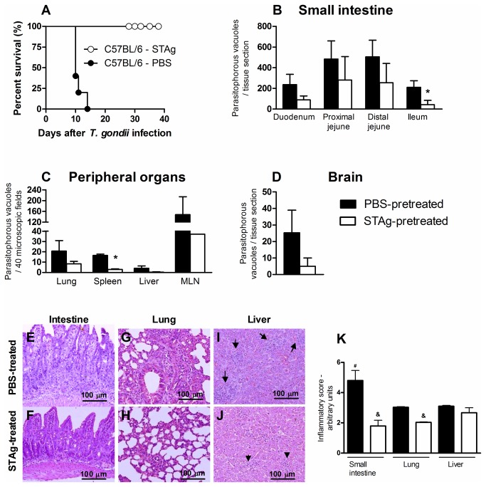
Figure 1. Mortality rates, tissue parasitism and inflammatory changes of STAg-pretreated C57BL/6 mice orally T. gondii infected.
The mortality rate for 8 mice from each group was determined (A). STAg-pretreated mice were significantly more resistant to toxoplasmosis than PBS-pretreated mice (χ2=10.03; P = .0015; df = 1). Tissue parasitism in the small intestine (B), peripheral organs (C) and brain (D) were detected on day 8 p.i. by immunohistochemistry staining and scored by counting the number of parasitophorous vacuoles per tissue section in the small intestine and brain and per 40 microscopic fields in the other peripheral organs, using a 40 x objective. The small intestine (E,F), lung (G,H) and liver (I,J) of PBS- and STAg-pretreated mice were stained by H&E and analyzed for histological changes. The inflammatory foci in the liver are shown (arrows). Bar scale, 100 µm. The data of inflammatory scores in the organs were obtained by analyzing 40 microscopic fields per section on six sections using a 40 x objective from each mouse (K). Data are representative of at least two independent experiments of 5 mice per group that provided similar results. *p < 0.05 (Significantly different from values obtained from PBS-pretreated mice, Unpaired Student’s t-test). &p < 0.03 (Significantly different from values obtained from PBS-pretreated mice, Mann Whitney test); # p < 0.05 (Significantly different from values obtained from lung and liver of PBS-pretreated mice, Kruskal-Wallis test and Dunn’s multiple comparisons post-test).

