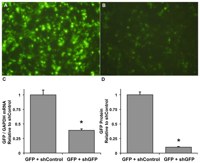
Figure 2. Efficient shRNA-mediated knockdown of GFP in vitro.
(A) AAV-293 cells dual-transfected with a plasmid expressing GFP from the cardiac troponin T promoter and a plasmid expressing a negative control shRNA from the mouse U6 promoter (shControl). (B) Cells transfected similarly but with the pAUSiGTL plasmid expressing shGFP. Cells were imaged 3 days post-transfection. (C) GFP mRNA from transfected cells harvested 3 days post-transfection. GAPDH was used to normalize GFP expression, which was found to be reduced by 61% compared to the shControl-treated group (n=3/group, p<0.01). (D) GFP protein expressed relative to the shControl-treated group showed a 91% reduction in the shGFP group (n=3/group, p<0.001).

