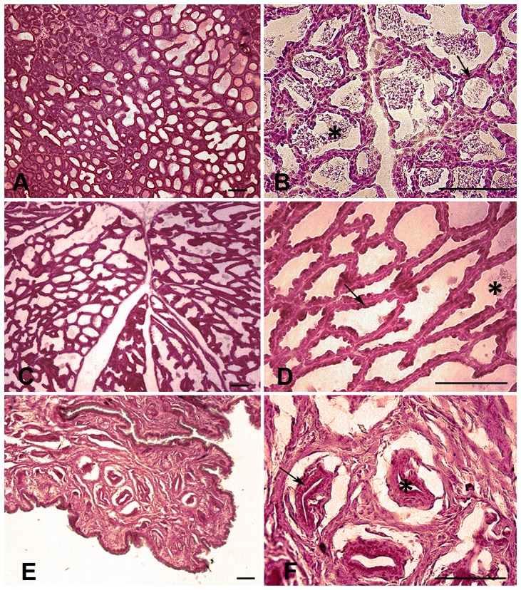
Figure 2. Histology of the mammary gland.

Photomicrographs showing the basic histology of the mammary gland of M. velifer in three stages: lactation (A and B) featuring secretion products inside the alveoli (asterisk) and epithelial alveolar cells (arrow); involution (C and D), with scarce secretion products inside the alveoli (asterisk) and epithelial alveolar cells (arrow); and the resting phase (E and F), where stroma occupies most of the gland (asterisk) and epithelial alveolar cells are found in a restricted area (arrow). Transverse sections. Scale: 100 µm.
