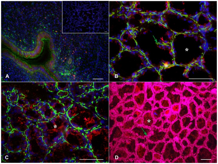
Figure 5. Localization of MAO A and TPH enzymes in myoepithelial and luminal epithelial cells.
Photomicrographs showing the localization of TPH and MAO A in luminal epithelial and myoepithelial cells of the mammary glands from lactating females. Figure 5A shows a section of rat uterus in estrus as a positive control and the negative control of mammary gland tissue in which incubation with primary antibody was omitted (inset). Figure 5B shows the co-localization of TPH (red, red arrow) in myoepithelial cells in yellow (green arrow), while Figure 5C shows the localization of MAO A (red arrow) and myoepithelial cells in green (green arrow). Figure 5D shows the co-localization of MAO A (purple arrow) in luminal epithelial cells in magenta (green arrow). Transversal sections. Scale: 100 µm.

