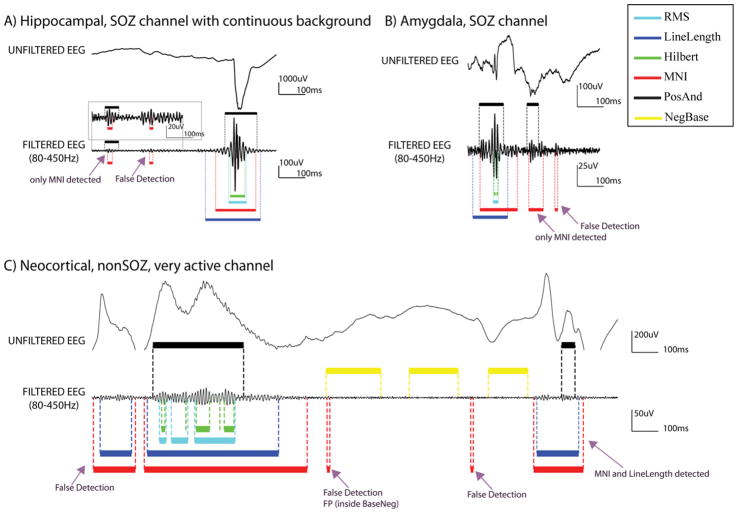
Fig. 6.
Examples of HFO detections. (A) SOZ channel in the hippocampus, with continuous background activity and many spikes as the one depicted in the raw EEG. All the detectors found the 2nd event, but only the MNI detector found also the 1st one. Baselines were not found, and therefore the detector for continuous channels block was used. In addition, a false detection is shown. (B) SOZ channel in the Amygdale. The 1st event was found by all detectors, the 2nd event only by the MNI detector. In this channel 10.6 s of baseline were found and HFOs were detected by the MNI detector based on that baseline. In addition, a false detection is shown. (C) NonSOZ, neocortical very active channel. The epileptiform activity that is observed is repeated periodically in the recording. In this channel 16.4 s of baseline were found. Therefore HFOs were detected by the MNI detector based on that baseline. Black: PosAnd reference HFOs; yellow: NegBase reference baseline; cyan: RMS detections; blue: LineLength detections; green Hilbert detections; and red: MNI detections. (For interpretation of the references in colour in this figure legend, the reader is referred to the web version of this article.)

