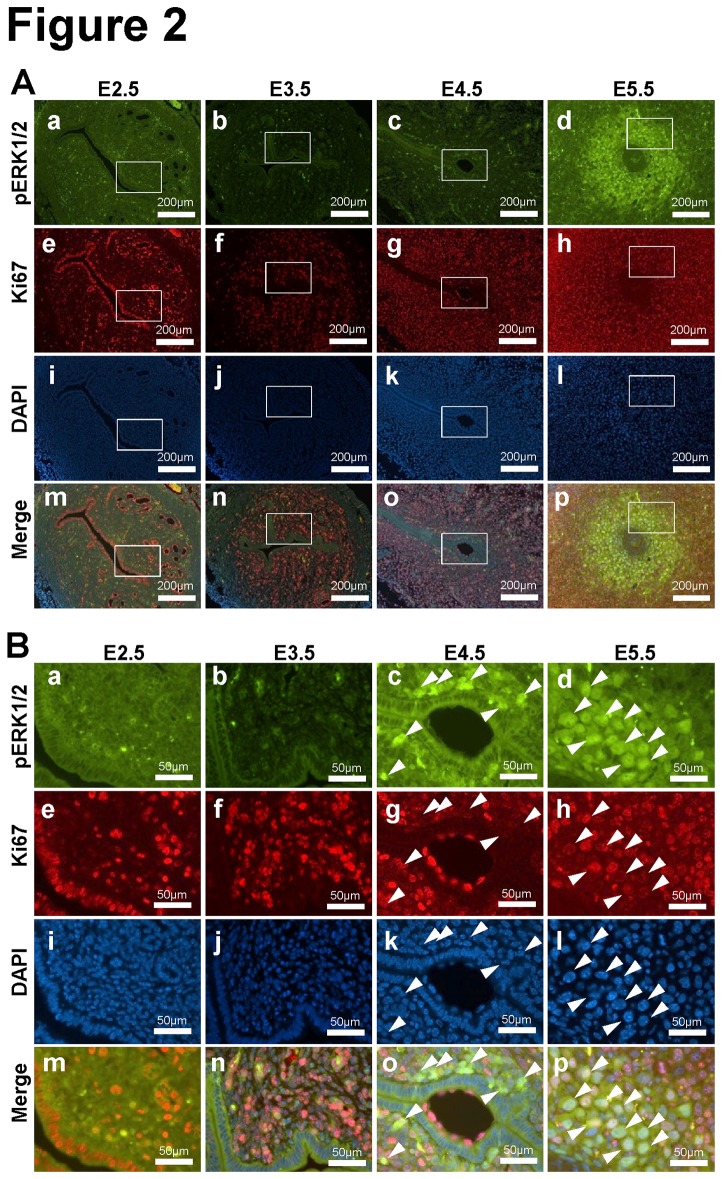
Figure 2. Relationship between phospho-ERK1/2 and proliferation during early pregnancy.
(A) Immunofluorescence analysis of phospho-ERK1/2 (green; a, b, c and d) and Ki67 (red: e, f, g and h) was performed in uteri of C57BL/6 mice on 2.5 (a, e, i and m), 3.5 (b, f, j and n), 4.5 (c, g, k and o) and 5.5 (d, h, l and p) dpc. Images (m, n, o and p) were merged with DAPI staining (i, j, k and l). (B) Higher magnification images of the insets. Arrowheads indicates positive phospho-ERK1/2 cells.

