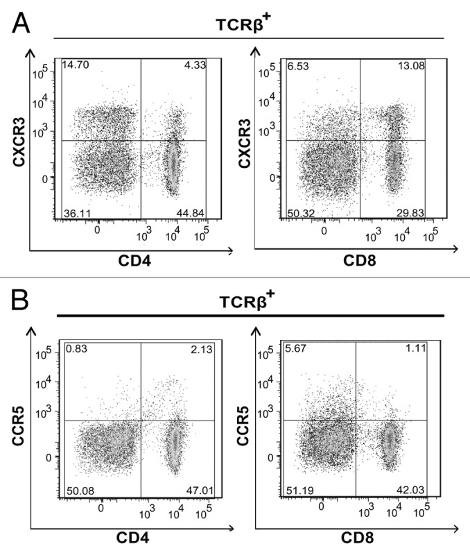
Figure 2. CD4+ and CD8+ T cells express CXCR3. (A and B) Lymph node (LN) cells were isolated from 3–5 tumor bearing mice that were vaccinated with live Lm-LLO-E7 bacteria, pooled and labeled with a panel of antibodies to identify multiple T-subsets. Labeled cells were analyzed by flow cytometry for the expression of CXCR3 and CCR5. Representative density plots are shown. CXCR3+ (A) and CCR5+ (B) T cells were identified among live, single TCRβ+ cells.
