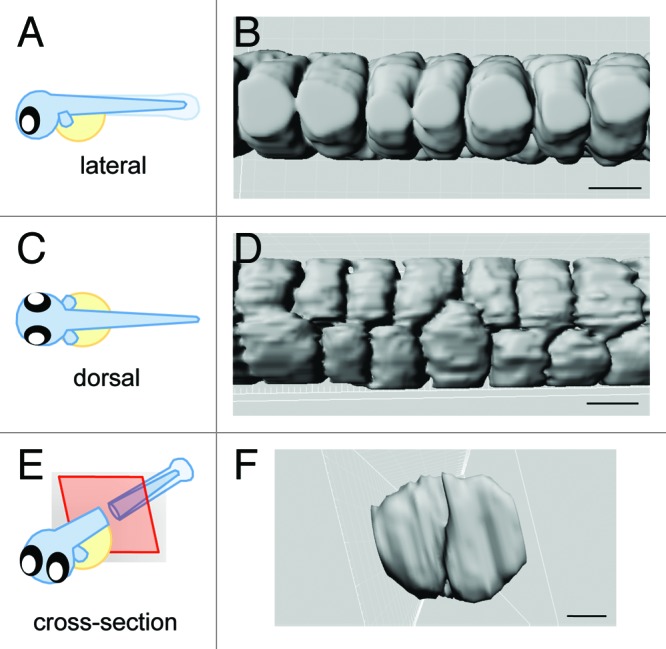
Figure 2. 3-Dimensional model of vacuolated cell arrangement. Imaris software was used to generate a 3-D model of vacuolated cells using a transgenic line that expresses GFP in the cytosol of the inner vacuolated cells, Gt(Gal4FF)nksagff214a; Tg(UAS:GFP).(A, C, E) Cartoons depicting orientation of the embryo for the following panels. (B, D, F) 3-D rendering of vacuolated cells. Scale bars = 30 µm.
