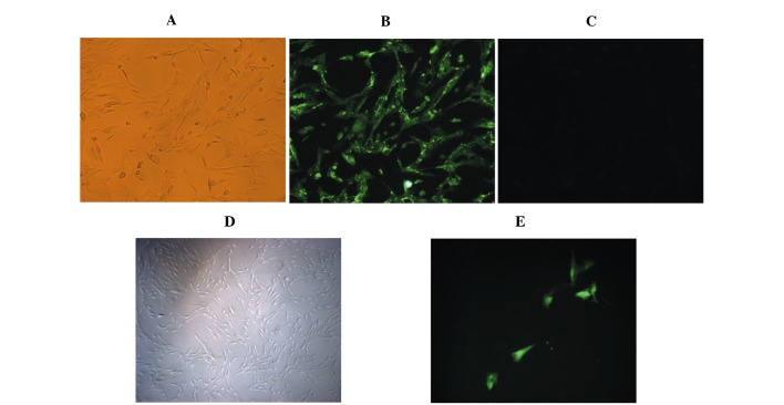
Figure 1.
(A and D) Cultured normal rat pulmonary microvascular endothelial cells (RPMVECs). (B) Positive group: RPMVECs were incubated with rabbit anti-rat CD31 antibody and FITC-labeled goat anti-rabbit IgG. (C) Negative group with 5% BSA instead of primary antibody incubation. (E) RPMVECs transfected with pcDNA3.1-eGFP and incubated for 48 h as the blank control group.

