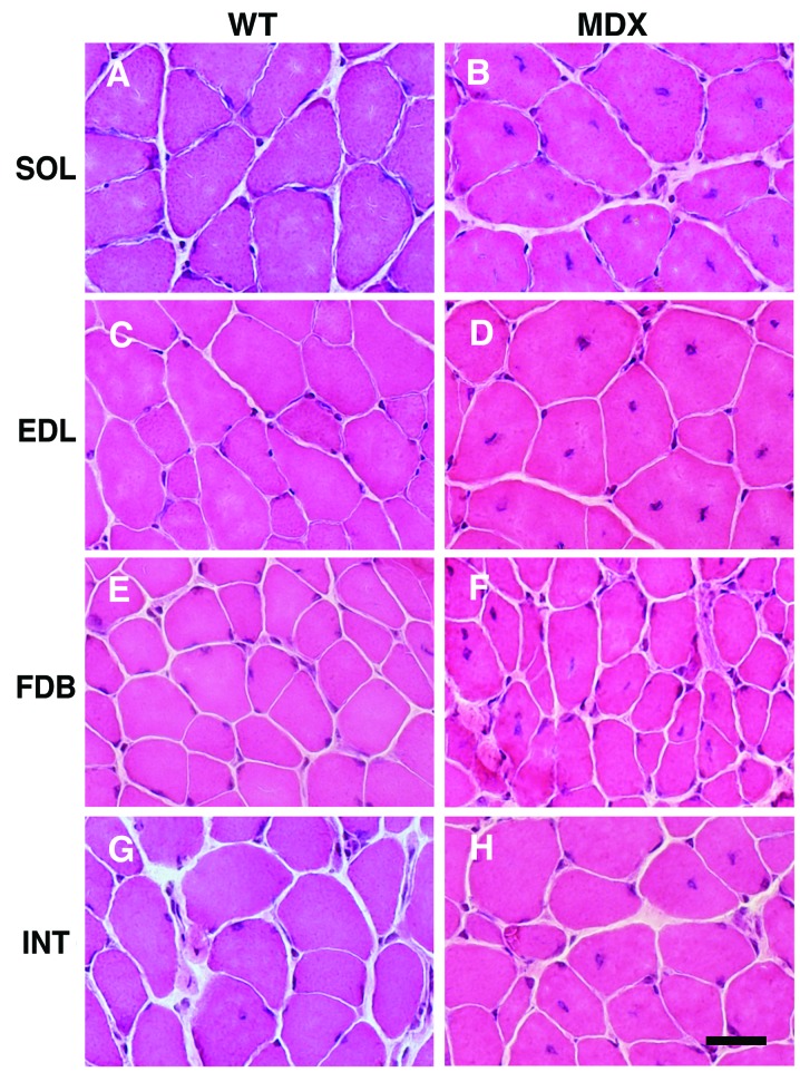
Figure 1.
Histological profiling of skeletal muscles from X-linked muscular dystrophy (mdx) mice. Shown are hematoxylin and eosin-stained muscle cross-sections from wild-type (WT; A, C, E and G) and mdx mice (B, D, F and H). Note the reduced occurrence of central nuclei in the flexor digitorum brevis (FDB) and interosseus (INT) muscles, compared with the soleus (SOL) muscle and extensor digitorum longus (EDL) muscle from mdx mice. Muscles were prepared from 100-day-old male animals. Scale bar, 20 μm.

