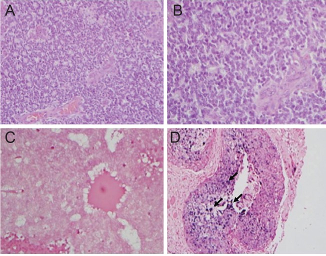
Fig. 1.

(A,B) Hematoxylin and eosin staining confirm the presence of retinoblastoma cells, ×200, ×400, respectively. (C) In situ hybridization for human papilloma virus (HPV) in retinoblastoma tumor cells is negative for HPV-DNA. (D) Squamous cell carcinoma of the uterine cervix stain positive for HPV-DNA. Dark purple staining (depicted in black arrows) indicates integrated or episomal HPV-DNA.
