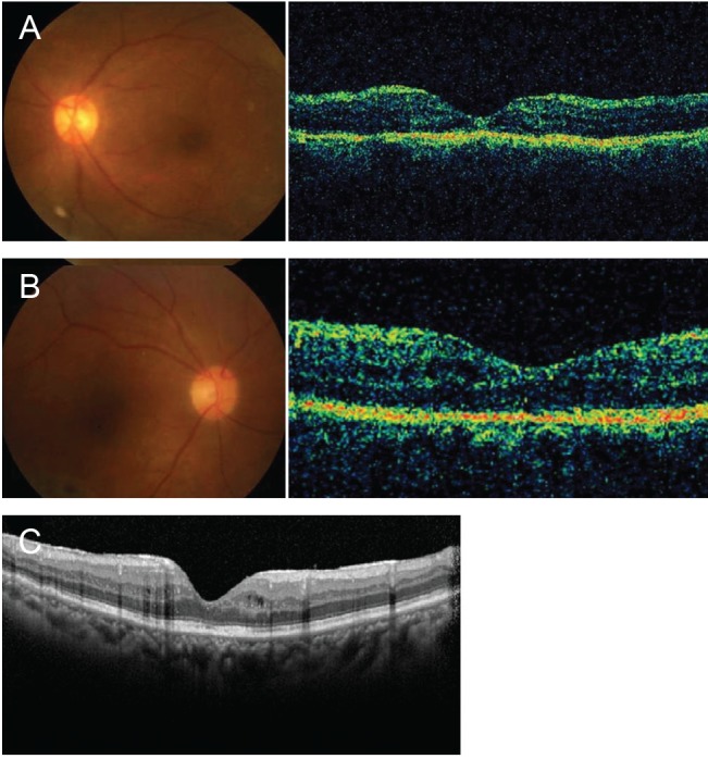
Fig. 4.

Fundus photograph (left) and macular optical coherence tomography (right) 2 weeks after surgery. (A) Case 1. (B) Case 2. Note that the subfoveal perfluorocarbon liquid (PFCL) bubbles and macular holes disappeared in both cases and that the retinal structures were better persevered in case 2. (C) Follow-up spectral-domain optical coherence tomography of case 2 at 1 year after PFCL removal shows the well-preserved retinal structures in the macula.
