Figure 4.
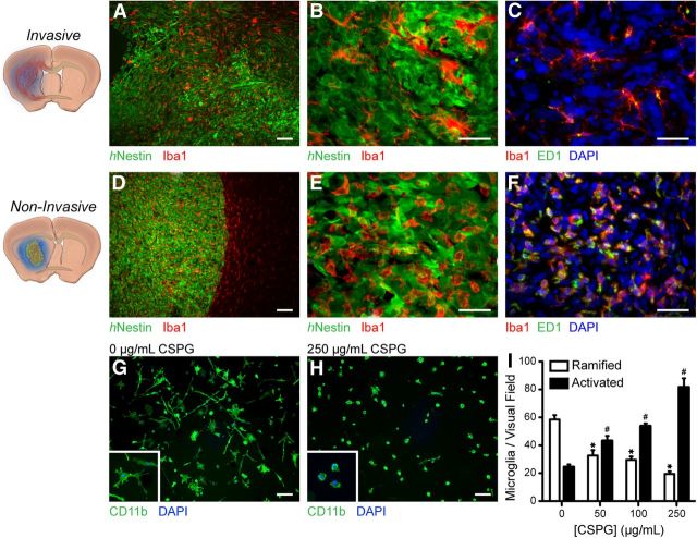
Microglial activation state differs markedly between invasive and noninvasive brain lesions. Representative low-magnification fluorescence micrographs demonstrate that microglia uniformly populate both (A) invasive and (D) noninvasive lesions. B, An elaborate ramified morphology and (C) weak ED-1 immune reactivity characterize invasive tumor-associated microglia. In comparison, microglia confined within CSPG-rich noninvasive lesions demonstrate (E) the amoeboid morphology and (F) intense ED-1 immune reactivity indicative of a heightened state of activation and chronic inflammation. G, inset, In vitro, in the absence of substrate-bound CSPGs, microglia maintain the ramified morphology indicative of the relatively decreased activation state observed in invasive tumor-associated microglia. H, inset, In contrast, microglia exposed to increasing concentrations of CSPGs match the activated, amoeboid morphology indicative of heightened activation as seen in noninvasive tumors. I, Quantification of microglial morphology demonstrates that CSPGs can serve as a potent activator of microglia (mean ± SEM, two-way ANOVA, Bonferroni-Dunn post hoc test, *p < 0.0001 as compared with 0, #p < 0.0001 as compared with 0, n = 30/group). Scale bars: A–F, 50 μm; G, H, 10 μm.

