Figure 1.
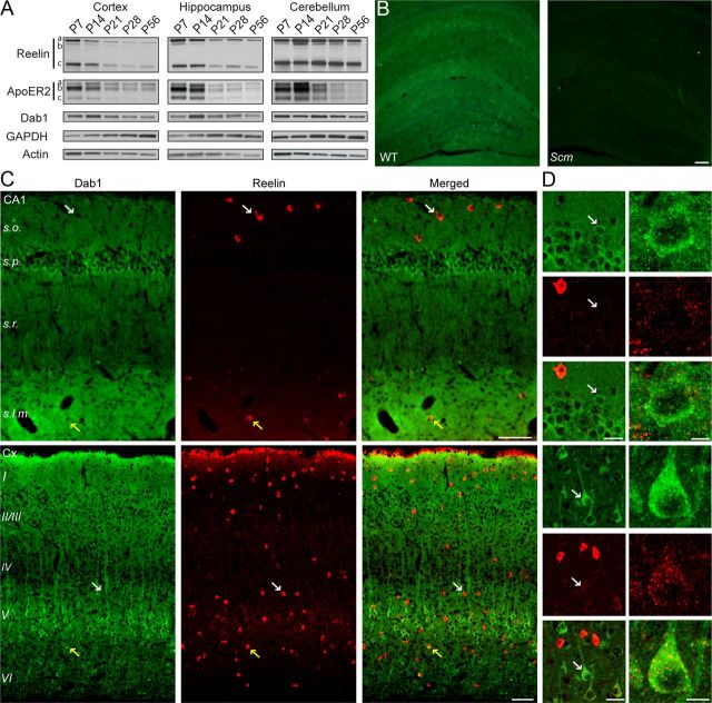
Dab1 and Reelin expression in the postnatal forebrain. A, Western blot analysis of Reelin, ApoER2, and Dab1 in the cortex, hippocampus, and cerebellum at P7, P14, P21, P28, and P56. Blots shown are representative of data obtained from 3 to 4 mice per age. Reelin was detected as a full-length isoform of ∼450 kDa (a) and two N-terminal fragments of 370 (b) and 180 kDa (c). ApoER2 was detected as three major isoforms (a, b, c). Dab1 was detected as a single band of ∼90 kDa. The blots were reprobed with GAPDH and actin antibodies as loading controls. B, Immunofluorescence labeling of Dab1 in the postnatal hippocampus. Dab1 signal (green) was present in WT examples, but not in the Dab1 mutant scrambler (Scm), confirming antibody specificity. Scale bar, 100 μm. C, D, Double immunofluorescence labeling of hippocampal area CA1 and the neocortex (Cx) with Dab1 (green) and Reelin (red) antibodies. Sections were obtained from 2- to 3-month-old WT mice. Larger panels show Dab1 and Reelin staining in mostly distinct cell populations (white arrows), with the exception of a few cells that showed colocalization (yellow arrows). D, Smaller panels show Dab1 labeling at higher magnification (left, white arrows). Scale bars: B and C, 100 μm; D, left, 20 μm; right, 10 μm.

