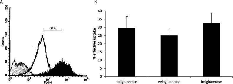
Figure 4. Uptake of β-glucocerebrosidase into U937 human macrophage like cell.
(A) U937 cells, treated with 75 ng/ml PMA, were stained with the macrophage marker CD11c. Unstained (negative control, grey), monocytes (untreated, open black) and macrophages (PMA treated, black) were analysed by flow cytometry. After differentiation, a 60% increase in CD11c staining can be seen; (B) PMA differentiated U937 macrophages were loaded with 60 μg/ml taliglucerase alfa, imiglucerase or velaglucerase alfa for 10 min at 37°C. The cells were then cooled and washed with acidic buffer. β-Glucocerebrosidase activity was tested in the cells before and after acid wash and the effective uptake is indicated. Results are the means±S.E.M. of six independent wells.

