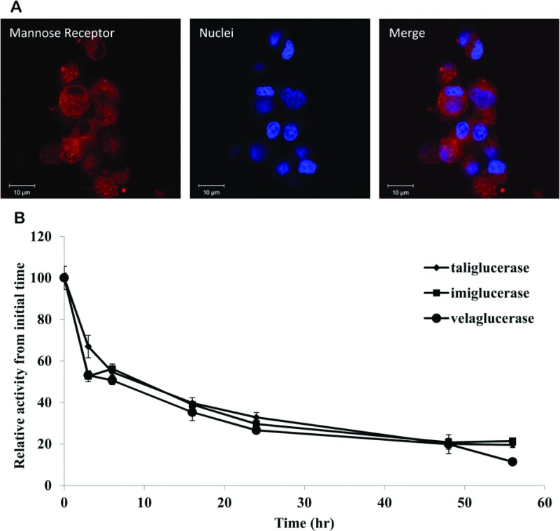
Figure 5. Uptake of β-glucocerebrosidase into rat alveolar macrophage cell line.
(A) Rat alveolar macrophage cell line NR8383 was fixed and stained with polyclonal anti-MR antibody produced in rabbit (1 mg/ml, Abcam), using donkey anti-rabbit conjugated to Cy3 (indocarbocyanine; Alexa Fluor) as the detection antibody. Nuclei were stained with DAPI. Pictures were taken by LSM Meta Confocal Microscopy, with a ×63 objective; (B) In-cell stability of taliglucerase alfa, imiglucerase or velaglucerase alfa. Enzymes were loaded into the cells at 60 μg/ml for two h. The cells were then washed and incubated up to 56 h. At each indicated time point, a sample was taken to assess the β-glucocerebrosidase activity within the cells. Results were normalized to the initial concentration of β-glucocerebrosidase in the cells. Results are means±S.E.M. of four independent wells.

