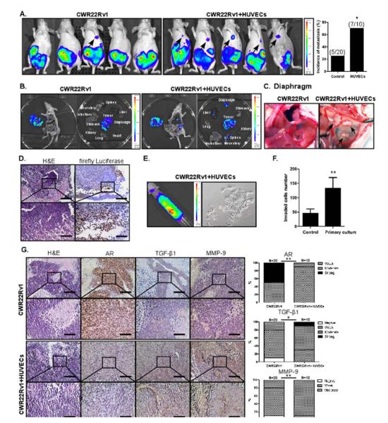
Fig. 6. HUVECs treatment enhances PCa metastasis in orthotopic xenografted mice.
CWR22Rv1 cells were transfected with luciferase (Luciferase-pcDNA3, Addgene), stable clones were selected, and their luciferase activity was confirmed before injection. 1 × 10⁶ of these cells, either alone or together with HUVECs (10:1 PCa cells:HUVECs), as a mixture with Matrigel, 1:1, total of 20 μl, were orthotopically implanted into the APs of 8 wks old mice. Tumor growth and metastasis was monitored by examining luminescence using IVIS at 3, 4, 5, and 6 wks after injection. (A) The metastatic incidence shown in two groups of mice. (B) The imaging data showing primary and metastatic tumors of two mice groups. (C) The imaging demonstrating diaphragm metastasis. (D) H&E and IHC staining of metastatic tumors from diaphragm using antibodies of anti-firefly Luciferase antibody (Abcam). (E) The imaging demonstrating the ascites metastases obtained from metastatic mouse (left panel) and primary cultures cells from ascites (right panel). (F) Invasion assay of primary cultured CWR22Rv1 from ascites (parental CWR22Rv1 cells as control). (G) H&E and IHC staining of primary and metastatic tumors using antibodies of AR, TGF-β1, and MMP-9.

