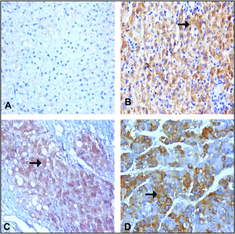
Figure 1.
Immune staining for TNF-α. (A) Normal hepatocytes from a control patient, showing negative expression of TNF-α in the cytoplasm of the hepatocytes. (B) CHC specimen without cirrhosis (A1F1) showing moderate expression of TNF-α in the cytoplasm of a hepatocyte (arrow). (C) A CHC specimen with cirrhosis (A2F3), showing a cirrhotic nodule with moderate to marked expression of TNF-α in the cytoplasm of a hepatocyte (arrow). (D) A case of moderately differentiated HCC, showing moderately expressed TNF-α in the cytoplasm of a hepatocyte (arrow). (A–D) IHC: DAB, ×200.

