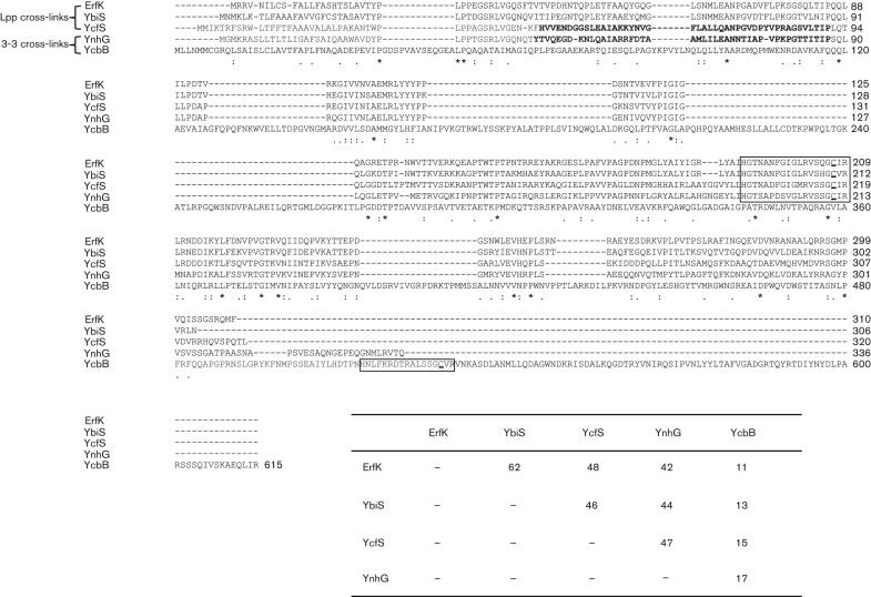
Fig. 1.
Clustal W (Larkin et al., 2007) protein sequence alignment of the five YkuD family Ldts from E. coli. The signature residues composing the catalytic domain found in all YkuD family proteins are boxed with the essential active site cysteine underlined. YcfS and YnhG both contain an N-terminal PG-binding LysM motif (bold). Lpp, lipoprotein. Percentage sequence similarity between the five proteins is shown in the inset.

