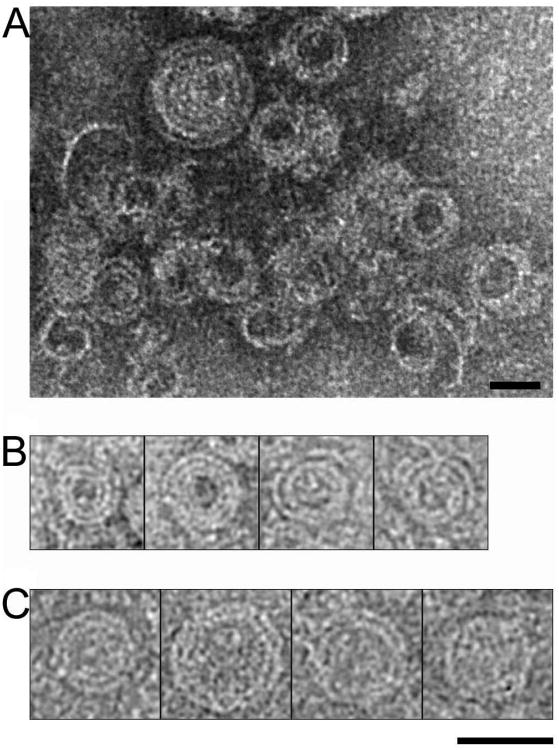
Figure 5. Conserved filament organization of ESCRT-III.
(A) Negative stained image of CHMP2AΔC polymers. (B) Gallery of spiral structures formed by CHMP2AΔC obtained by negative staining electron microscopy and (C) cryo-EM. CHMP2AΔC forms 30 Å-thick strands, which are similar to the width of CHMP2B and CHMP2AΔC-CHMP3 filaments. Scale bars are 400 Å.

