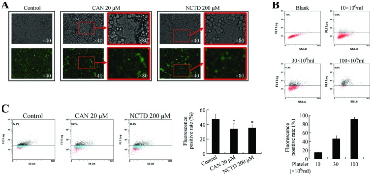
Figure 2.
(A) Photomicrographs and fluorescence microscopy images of adhesion between MCF-7 cells and platelets. (B) Flow cytometry-based platelet adhesion assay. The fluorescent positive rate increased when the platelet concentration increased. (C) Cantharidin (CAN) and norcantharidin (NCTD) treatment decreased the fluorescent positive rate. *P<0.05 vs. control group.

