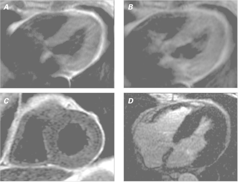
Fig. 3 Cardiac magnetic resonance with a myocarditis protocol. Spin-echo T1-weighted images show increased global early myocardial enhancement A) before and B) after gadolinium contrast, yielding an elevated ratio of 6.1 (normal, <4). C) Fast-spin-echo T2-weighted image shows global myocardial edema with an elevated ratio (2.1) of T2 signal in the myocardium compared to skeletal muscle (normal, <2). D) Late-gadolinium-enhancement image shows no focal myocardial fibrosis or injury.

An official website of the United States government
Here's how you know
Official websites use .gov
A
.gov website belongs to an official
government organization in the United States.
Secure .gov websites use HTTPS
A lock (
) or https:// means you've safely
connected to the .gov website. Share sensitive
information only on official, secure websites.
