Figure 5.
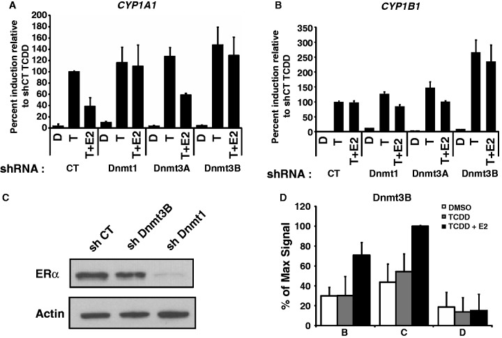
ERα can not repress CYP1A1 induction in Dnmt3B-depleted cells. CYP1A1 (A) and CYP1B1 (B) expression was measured in MCF7 cells infected with shCT, shDnmt1, shDnmt3A or shDnmt3B constructs for 5 days and then treated with DMSO (D), 10 nM TCDD (T) or 10 nM TCDD + 100 nM E2 (T+E2) for 24 h. (C) MCF7 cells were infected with shCT, shDnmt1 and shDnmt3b constructs for 5 days, and then proteins were extracted and western blot performed to verify ERα protein levels. Actin is used as loading control. ChIP of Dnmt3B (D) was performed in MCF7 cells grown in estrogen-free media for 3 days, and then treated with DMSO, TCDD or TCDD + E2 for 90 min.

